Tillie Brandes
Tillie Brandes
  • Blog
  • About
  • Contact

Best Ways To Spot Calcaneal Spur

9/26/2015

 
Posterior Calcaneal Spur

Overview

Heel spurs are a common foot problem resulting from excess bone growth on the heel bone. The bone growth is usually located on the underside of the heel bone, extending forward to the toes. One explanation for this excess production of bone is a painful tearing of the plantar fascia connected between the toes and heel. This can result in either a heel spur or an inflammation of the plantar fascia, medically termed plantar fascitis. Because this condition is often correlated to a decrease in the arch of the foot, it is more prevalent after the age of six to eight years, when the arch is fully developed.

Causes

Causes of Heel Spur Syndrome are mostly due to your foot structure. Foot structure is due to hereditary for the most part, meaning it was the way you were when born. Other factors such as increase in weight, injury, improper shoes, or different activities may change the way your foot functions as well. If one leg is longer or shorter than the other, this may make your foot function improperly and be the cause of the heel spur syndrome. Improper shoes may be ones that are new or ones that are worn out and do not give good support. The higher priced shoes do not mean it's a better shoe. Pronation is a term used to describe a foot which allows the arch to fall more than normal and allows for the fascia along the bottom of the foot to put a tighter pull or a different angle of pull on the heel bone. Over time, this constant pull of the tight fascia can force the bone to enlarge and form a spur. It is not the heel spur that causes the pain directly. The spur may cause pressure against a nearby nerve causing a neuritis, or a bursa causing a bursitis.

Posterior Calcaneal Spur

Symptoms

Most bone spurs cause no signs or symptoms. You might not realize you have bone spurs until an X-ray for another condition reveals the growths. In some cases, though, bone spurs can cause pain and loss of motion in your joints.

Diagnosis

Sharp pain localized to the heel may be all a doctor needs to understand in order to diagnose the presence of heel spurs. However, you may also be sent to a radiologist for X-rays to confirm the presence of heel spurs.

Non Surgical Treatment

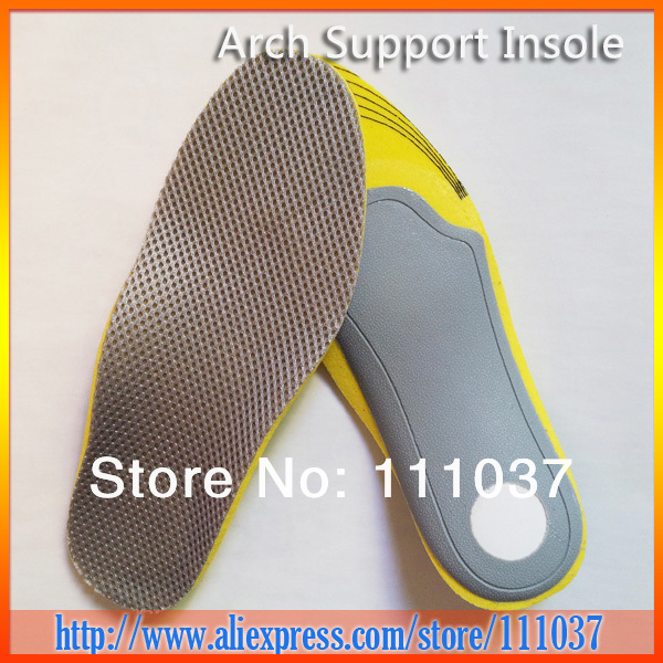
In extreme cases, a doctor may recommend surgery for the removal of heel spurs. Fortunately, this is the exception rather than the rule. Most cases can be resolved with a combination of icing, rest, foot stretches and supporting the foot with an orthodic shoe insert specifically designed for this condition. We recommend that you continue on to our article on Heel Spur Treatment to discover the best, speediest and most affordable methods of resolving this ailment without invasive medical procedures.

Surgical Treatment

In a small number of cases (usually less than 5 percent), patients may not experience relief after trying the recommendations listed above. It is important that conservative treatments (such as those listed above) be performed for AT LEAST a year before considering surgery. Time is important in curing the pain from heel spurs, and insufficient treatment before surgery may subject you to potential complications from the procedure. If these treatments fail, your doctor may consider an operation to loosen the plantar fascia, called a plantar fascia release.

Bursitis Of The Foot Pain Therapy

8/23/2015

 
Overview

Heel bursitis is a common foot pain in athletes, and is often mistaken for Achilles tendinitis, although it can also occur together with Achilles tendinitis. Heel bursitis occurs when small cushions in the heel called bursas become inflamed and swell with fluid irritating surrounding tissue and pressing on nerves. Other names given to heel bursitis are Achilles bursitis and Retrocalcaneal bursitis.

Causes

Retrocalcaneal bursitis is generally caused by local trauma from poorly designed shoes. Patients complain of posterolateral heel pain and may have a posterior heel prominence (?pump bump?), as well as local swelling and tenderness over the Achilles tendon. Pain is increased by squeezing the bursa from side to side and anterior to the Achilles. A heel lift and open-back shoes help alleviate pressure.

Symptoms

Limping. Decreased movement. Your ankles may feel stiff or unable to move as well as they usually do. Pain or tenderness in the back of the ankle. It may be worse at the beginning of exercise, or when running uphill. You may also have pain when wearing shoes. Redness and warmth. If the bursa is infected, the skin over the heel may be red and warm. You may also have a fever. Swelling on the back of the heel.

Diagnosis

Your health care provider will take a history to find out if you have symptoms of retrocalcaneal bursitis. Examining your ankle can find the location of the pain. The physician will look for tenderness and redness in the back of the heel. The pain may be worse when the doctor bends the ankle upward (dorsiflex). Or, the pain may be worse when you rise on your toes. You will not usually need imaging studies such as x-ray and MRI at first. If the first treatment does not improve the symptoms, your health care provider may recommend these tests. MRI may show inflammation.

Non Surgical Treatment

Non-operative treatment is the standard approach to treating posterior heel pain. It is highly desirable to treat this condition non-operatively, as operative treatment is often associated with a prolonged recovery. Traditional non-operative treatment includes the following. Heel Lift or the Use of a Shoe with a Moderate Heel. Walking barefoot or in a flat-soled shoe increases the tension on the insertion of the Achilles tendon. Using a heel lift or a shoe with a moderate heel can help reduce the stress on the tendon and decrease the irritation caused by this condition.

Surgical Treatment

Surgery is rarely need to treat most of these conditions. A patient with a soft tissue rheumatic syndrome may need surgery, however, if problems persist and other treatment methods do not help symptoms.

Recovery After Bunion Hammer Toe Surgery

6/23/2015

 
HammertoeOverview

A hammertoe is a toe that tends to remain bent at the middle joint in a claw-like position. There are 2 types of hammer toe. Flexible hammer toe, can be straightened by hand. Rigid hammer toe, cannot be pulled straight and can be extremely painful. The position of the toe can also lead to corns or calluses. These may also be painful. Hammer toe may be present at birth or develop later in life due to tendons that have tightened, causing the toe's joints to curl downward. Occasionally, all toes may be bent. This may be due to problems with the peripheral nerves or the spinal cord.

Causes

Hammer toe usually affects the second toe. However, it may also affect the other toes. The toe moves into a claw-like position. The most common cause of hammer toe is wearing short, narrow shoes that are too tight. The toe is forced into a bent position. Muscles and tendons in the toe tighten and become shorter. Hammer toe is more likely to occur in women who Hammer toes wear shoes that do not fit well or have high heels and children who keep wearing shoes they have outgrown. The condition may be present at birth (congenital) or develop over time. In rare cases, all of the toes are affected. This may be caused by a problem with the nerves or spinal cord.

Hammer ToeSymptoms

Patients with hammer toe(s) may develop pain on the top of the toe(s), tip of the toe, and/or on the ball of the foot. Excessive pressure from shoes may result in the formation of a hardened portion of skin (corn or callus) on the knuckle and/or ball of the foot. Some people may not recognize that they have a hammer toe, rather they identity the excess skin build-up of a corn.The toe(s) may become irritated, red, warm, and/or swollen. The pain may be dull and mild or severe and sharp. Pain is often made worse by shoes, especially shoes that crowd the toes. While some hammer toes may result in significant pain, others may not be painful at all. Painful toes can prevent you from wearing stylish shoes.

Diagnosis

First push up on the bottom of the metatarsal head associated with the affected toe and see if the toe straightens out. If it does, then an orthotic could correct the problem, usually with a metatarsal pad. If the toe does not straighten out when the metatarsal head is pushed up, then that indicates that contracture in the capsule and ligaments (capsule contracts because the joint was in the wrong position for too long) of the MTP joint has set in and surgery is required. Orthotics are generally required post-surgically.

Non Surgical Treatment

Treatment for a hammertoe usually depends on the stage of the hammertoe and the cause of the condition. If your toe is still bendable, your doctor may suggest conservative care-relieving pressure with padding and strapping, or proper shoes that have a deep toe box and are of adequate length and width. Early intervention can often prevent the need for surgery.

Surgical Treatment

Surgery may not help how your foot looks. And your toe problems may also come back after surgery. This is more likely if you keep wearing the kinds of shoes that cause toe problems. Your expectations will play a large role in how you feel about the results of surgery. If you are only having surgery to improve the way your foot looks, you may not be happy with how it turns out.

The Truth About Overpronation Of The Feet

6/2/2015

 
Overview

Too much of a good thing can be bad, and when it comes to pronation of the foot this is often the case. When the foot rolls too much during contact with the ground, the body weight is not cushioned effectively. The degree to which pronation occurs may be difficult to spot without someone observing how you run. It is easier to tell if a runner is pronating excessively when they are observed from behind rather than from above. When overpronation is only mild to moderate it may not be easy to spot without having a professional gait analysis performed.Over Pronation

Causes

Congenital "Flat Feet" - an individual may be born with feet that lack an appropriately supportive arch thereby predisposing the individual to this foot condition. Excessive Weight (Obesity) Too much weight on the foot from either obesity or pregnancy may be a factor. Repetitive Impact walking on flat, hard surfaces continuously places unnatural stress on the foot arch.

Symptoms

Overpronation can negatively affect overall body alignment. The lowering of the longitudinal arch pulls the heel bone in, causing the leg, thigh bone and hip to rotate inwards, and an anterior tilt of the pelvis. Unnecessary strain to the ankles, knees, hips and back can result. Plantar fasciitis and inflammation, metatarsal pain, problems with the Achilles tendon, pain on the inside of the knee, and bursitis in the hip are just some of the conditions commonly associated with pronation.

Diagnosis

Look at your soles of your footwear: Your sneaker/shoes will display heavy wear marks on the outside portion of the heel and the inside portion above the arch up to the top of the big toe on the sole. The "wet-foot" test is another assessment. Dip the bottom of your foot in water and step on to a piece of paper (brown paper bag works well). Look at the shape of your foot. If you have a lot of trouble creating an arch, you likely overpronate. An evaluation from a professional could verify your foot type.Pronation

Non Surgical Treatment

The following exercises help retrain the foot and ankle complex to correct overpronation. Exercises may be performed while wearing shoes, or for an even greater challenge, in bare feet. Duck Stand. This exercise is designed to prepare for the more dynamic BT exercises ahead by waking up the gluteal muscles and teaching clients how the gluteal muscles control the degree of foot pronation. For example, when the glutes contract concentrically, they rotate the leg outward. As the leg rotates outward, the arch of the foot raises (i.e., supinates). Stand beside the BT with both heels together and feet turned outward. (Note: As you progress, perform this exercise while standing on the BT.) Try to rotate legs outward by tightening buttock muscles while tilting pelvis under. As legs rotate outward, arches of the feet raise up out of pronation. Hold position for 30 seconds. Big Toe Pushdowns. This exercise is designed to strengthen the muscle of the big toe that holds up the arch of the foot (i.e., flexor hallucis longus muscle). This stops the foot from overpronating. Stand on top of the BT dome with feet facing forward. Use gluteal muscles to raise the arches of the feet (see previous exercise - "Duck Stand"). Keep arches raised while pushing down big toe into the BT. While pushing down, tension build in the arch on the underside of their foot should be felt. Hold position for 15 seconds.

Surgical Treatment

Hyperpronation can only be properly corrected by internally stabilizing the ankle bone on the hindfoot bones. Several options are available. Extra-Osseous TaloTarsal Stabilization (EOTTS) There are two types of EOTTS procedures. Both are minimally invasive with no cutting or screwing into bone, and therefore have relatively short recovery times. Both are fully reversible should complications arise, such as intolerance to the correction or prolonged pain. However, the risks/benefits and potential candidates vary. Subtalar Arthroereisis. An implant is pushed into the foot to block the excessive motion of the ankle bone. Generally only used in pediatric patients and in combination with other procedures, such as tendon lengthening. Reported removal rates vary from 38% - 100%, depending on manufacturer. HyProCure Implant. A stent is placed into a naturally occurring space between the ankle bone and the heel bone/midfoot bone. The stent realigns the surfaces of the bones, allowing normal joint function. Generally tolerated in both pediatric and adult patients, with or without adjunct soft tissue procedures. Reported removal rates, published in scientific journals vary from 1%-6%.

Learn How To Spot Severs Disease?

5/20/2015

 
Overview

Sever's disease involves a lesion to the heel that appears mostly as a result of excessively twisting the ankle. The injury is particularly prevalent in children over 10 and other younger athletes because the heel bone, muscles and tendons are still developing - the bone faster than the muscles - and the condition strikes a growing part of the bone called the calcaneal apophysis. In older people an injury of this sort generally results in a stress fracture.

Causes

There are several factors which may increase the likelihood of developing this condition. These need to be assessed and corrected with direction from a physiotherapist to ensure an optimal outcome. Some of these factors include inappropriate footwear, calf tightness and/or weakness, joint stiffness (particularly the foot and ankle), poor lower limb biomechanics, inappropriate or excessive training, inadequate recovery periods from training or activity, inappropriate training surfaces, inadequate warm up, poor core stability, a lack of lower limb strength and stability, poor proprioception or balance, rapid growth and age.

Symptoms

The most prominent symptom of Sever's disease is heel pain which is usually aggravated by physical activity such as walking, running or jumping. The pain is localized to the posterior and plantar side of the heel over the calcaneal apophysis. Sometimes, the pain may be so severe that it may cause limping and interfere with physical performance in sports. External appearance of the heel is almost always normal, and signs of local disease such as edema, erythema (redness) is absent. The main diagnostic tool is pain on medial- lateral compression of the calcaneus in the area of growth plate, so called squeeze test. Foot radiographs are usually normal. Therefore the diagnosis of Sever's disease is primarily clinical.

Diagnosis

Sever's disease is diagnosed based on a doctor?s physical examination of the lower leg, ankle, and foot. If the diagnosis is in question, the doctor may order X-rays or an MRI to determine if there are other injuries that may be causing the heel pain.

Non Surgical Treatment

Sever?s disease is believed to be the byproduct of repepitive stress and trauma to the growth plate in the calcaneous, or heel bone. The stress is from the pull of the Achilles tendon on the growth plate where the tendon attaches.

Exercise

The following exercises are commonly prescribed to patients with Severs disease. You should discuss the suitability of these exercises with your physiotherapist prior to beginning them. Generally, they should be performed 1 - 3 times daily and only provided they do not cause or increase symptoms. Your physiotherapist can advise when it is appropriate to begin the initial exercises and eventually progress to the intermediate, advanced and other exercises. As a general rule, addition of exercises or progression to more advanced exercises should take place provided there is no increase in symptoms. Calf Stretch with Towel. Begin this stretch in long sitting with your leg to be stretched in front of you. Your knee and back should be straight and a towel or rigid band placed around your foot as demonstrated. Using your foot, ankle and the towel, bring your toes towards your head as far as you can go without pain and provided you feel no more than a mild to moderate stretch in the back of your calf, Achilles tendon or leg. Hold for 5 seconds and repeat 10 times at a mild to moderate stretch provided the exercise is pain free. Calf Stretch with Towel. Begin this exercise with a resistance band around your foot and your foot and ankle held up towards your head. Slowly move your foot and ankle down against the resistance band as far as possible and comfortable without pain, tightening your calf muscle. Very slowly return back to the starting position. Repeat 10 - 20 times provided the exercise is pain free. Once you can perform 20 repetitions consistently without pain, the exercise can be progressed by gradually increasing the resistance of the band provided there is no increase in symptoms. Bridging. Begin this exercise lying on your back in the position demonstrated. Slowly lift your bottom pushing through your feet, until your knees, hips and shoulders are in a straight line. Tighten your bottom muscles (gluteals) as you do this. Hold for 2 seconds then slowly lower your bottom back down. Repeat 10 times provided the exercise is pain free.

Flat Foot Symptoms In Adults

4/19/2015

 
Overview
Flatfoot deformity is a general term used to describe a person whose arches are slowly dropping to the ground, aka ?fallen arches.? Adult-acquired flatfoot deformity can be caused by several factors, but the most common is abnormal functioning of the posterior tibial tendon in the foot and ankle. The posterior tibial tendon is the primary tendon that supports the arch. If this tendon begins to elongate from a sustained, gradual stretch over a long period of time, then the arch will progressively decrease until full collapse of the arch is noted on standing. What makes this tendon elongated? Biomechanical instability of the foot such as over-pronation or an accessory bone at the insertion site of the tendon are the primary causes for posterior tibial tendon dysfunction. Acquired Flat Foot

Causes
Rheumatoid arthritis This type of arthritis attacks the cartilage in the foot, leading to pain and flat feet. It is caused by auto-immune disease, where the body?s immune system attacks its own tissues. Diabetes. Having diabetes can cause nerve damage and affect the feeling in your feet and cause arch collapse. Bones can also fracture but some patients may not feel any pain due to the nerve damage. Obesity and/or hypertension (high blood pressure) This increases your risk of tendon damage and resulting flat foot.

Symptoms
Symptoms of pain may have developed gradually as result of overuse or they may be traced to one minor injury. Typically, the pain localizes to the inside (medial) aspect of the ankle, under the medial malleolus. However, some patients will also experience pain over the outside (lateral) aspect of the hindfoot because of the displacement of the calcaneus impinging with the lateral malleolus. This usually occurs later in the course of the condition. Patients may walk with a limp or in advanced cases be disabled due to pain. They may also have noticed worsening of their flatfoot deformity.

Diagnosis
Looking at the patient when they stand will usually demonstrate a flatfoot deformity (marked flattening of the medial longitudinal arch). The front part of the foot (forefoot) is often splayed out to the side. This leads to the presence of a ?too many toes? sign. This sign is present when the toes can be seen from directly behind the patient. The gait is often somewhat flatfooted as the patient has the dysfunctional posterior tibial tendon can no longer stabilize the arch of the foot. The physician?s touch will often demonstrate tenderness and sometimes swelling over the inside of the ankle just below the bony prominence (the medial malleolus). There may also be pain in the outside aspect of the ankle. This pain originates from impingement or compression of two tendons between the outside ankle bone (fibula) and the heel bone (calcaneus) when the patient is standing.

Non surgical Treatment
Nonoperative treatment of posterior tibial tendon dysfunction can be successful with the Arizona AFO brace, particularly when treatment is initiated in the early stages of the disease. This mandates that the orthopedist has a high index of suspicion when evaluating patients to make an accurate diagnosis. Although there is a role for surgical management of acquired flat feet, a well-fitted, custom-molded leather and polypropylene orthosis can be effective at relieving symptoms and either obviating or delaying any surgical intervention. In today's climate of patient satisfaction directed health care, a less invasive treatment modality that relieves pain may prove to be more valuable than similar pain relief that is obtained after surgery. Questions regarding the long-term results of bracing remain unanswered. Future studies are needed to determine if disease progression and arthrosis occur despite symptomatic relief with a brace. Furthermore, age- and disease stage-matched control groups who are randomized to undergo surgery or bracing are necessary to compare these different treatment modalities. Adult Acquired Flat Feet

Surgical Treatment
Surgery should only be done if the pain does not get better after a few months of conservative treatment. The type of surgery depends on the stage of the PTTD disease. It it also dictated by where tendonitis is located and how much the tendon is damaged. Surgical reconstruction can be extremely complex. Some of the common surgeries include. Tenosynovectomy, removing the inflamed tendon sheath around the PTT. Tendon Transfer, to augment the function of the diseased posterior tibial tendon with a neighbouring tendon. Calcaneo-osteotomy, sometimes the heel bone needs to be corrected to get a better heel bone alignment. Fusion of the Joints, if osteoarthritis of the foot has set in, fusion of the joints may be necessary.

Pain In The Arches Cause And Effect

4/14/2015

 
Overview

A fallen arch or flatfoot is known medically as pes planus. The foot loses the gently curving arch on the inner side of the sole, just in front of the heel. If this arch is flattened only when standing and returns when the foot is lifted off the ground, the condition is called flexible pes planus or flexible flatfoot. If the arch disappears in both foot positions, standing and elevated, the condition is called rigid pes planus or rigid flatfoot.

Arch Pain

Causes

A common cause of foot arch pain is a stress fracture. They tend to occur from repeated overloading of one of the foot bones from activities such as jumping and running especially if you have suddenly increased your activity level. The breaks in the bone may be small but they can be extremely painful. Stress fractures of the metatarsal bones or the navicular can cause anything from mild to severe foot arch pain. The Tibialis Posterior muscle plays a very important role in supporting the medial arch of the foot. Posterior Tibial Tendonitis can occur either through repetitive use e.g. high impact sports such as soccer or tennis, or from an injury e.g. a fall. This causes the tendon to become inflamed or even torn, resulting in pain on bottom of foot. This pain usually gets worse with activity or when standing for long periods. If the problem persists, the inner side of the foot (known as the medial longitudinal arch of the foot) gradually collapses down, causing flat feet. A simple test for this condition is to stand on one leg and rise up onto your tiptoes. If you cannot, it indicates a problem with the Posterior Tibial tendon. Treatment usually consists of rest, ice, exercises, orthotics and physical therapy.

Symptoms

Common symptoms of plantar fasciitis include pain in the morning when you first get out of bed, pain and stiffness when you start to walk after sitting for a while, increasing arch or heel pain toward the end of the day, tired feet at the end of the day. Other causes of arch and heel pain include arthritis, infection, fractures and sprains, and even certain systemic diseases. Since there are multiple possible causes, you should see your podiatrist for a thorough evaluation if you are experiencing arch or heel pain that does not respond quickly to early treatment.

Diagnosis

Diagnosis of a plantar plate tear can often be challenging due to the complex nature of the anatomy of the foot. Careful history taking and an examination of the area of pain is required to determine the extent and cause of the tear. If necessary, further investigations such as x-rays or diagnostic ultrasound may be ordered by your podiatrist to help evaluate the severity of the problem.

Non Surgical Treatment

Consult a doctor to diagnose the condition and determine the cause. If revealed to be plantar fasciitis, please refer to our article on that injury for further information. Generally arch pain is easy to treat, with the most effective method of treatment being the placement of arch supports in the shoes. This counteracts the strain placed on the arches by biomechanical errors, causing them to cease stretching excessively. A specialist can recommend the inserts suitable to your needs, which will depend on the shape of your arches. These supports should lessen your symptoms within days. If pain is severe you should refrain from running activities until it subsides to avoid risking an aggravation of the injury. To maintain fitness, alter your training program temporarily to be focused on low-impact sports like swimming. Applying ice to the affected area should assist in reducing pain and swelling.

Pain In Arch

Surgical Treatment

In rare cases, surgery may be needed if a child has flat feet caused by a problem they're born with (a congenital abnormality). The foot may need to be straightened or the bones may need to be separated if they're fused together. Painkillers and insoles are the first treatment options for flat feet that are caused by a joint problem, such as arthritis or a torn tendon. However, surgery may be recommended if the injury or condition is severely affecting your feet. Where flat feet are caused by a condition that affects the nervous system, special shoes, insoles, or supportive foot or leg braces may be needed. Again, in severe cases, an operation may be needed to straighten the feet.

Prevention

Foot and ankle injuries are common in sports, especially running, tennis and soccer. But sports enthusiasts can decrease the risk of injury by taking some precautions. Lightly stretch or better yet, do a slow jog for two to three minutes to warm up the muscles. Don't force the stretch with a "bouncing motion." The amount of time spent on the activity should be increased gradually over a period of weeks to build both muscle strength and mobility. Cross training by participating in different activities can help build the muscles. People whose feet pronate or who have low arches should choose shoes that provide support in both the front of the shoe and under the arch. The heel and heel counter (back of the shoe) should be very stable. Those with a stiffer foot or high arches should choose shoes with more cushion and a softer platform. Use sport-specific shoes. Cross training shoes are an overall good choice; however, it is best to use shoes designed for the sport.

Stretching Exercises

Strength training and stretching can help avoid injury and keep your feet free from pain. Stretching should focus on the bottom of your foot to loosen tissues and tight ligaments surrounding your arch. The easiest way to do this is by grabbing a towel and sitting on the floor. You can do this while you catch up on the news in the morning, or when you get home from work. Put one leg out in front with your foot flexed up. Loop the towel around the ball of your foot and gently pull your toes towards you. Hold for thirty seconds and then repeat 3-4 times before switching feet.

Achilles Tendon Sheath Problems

4/4/2015

 
Overview

Achilles Tendon
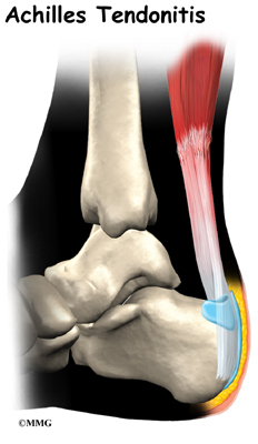
The Achilles tendon attaches the calf muscle to the heel bone. Jumping, climbing and strenuous exercise can strain the tendon and calf muscle, which can cause the type of inflammation known as tendinitis. This injury can be mild enough that it can be treated by over-the-counter medications or so severe that it must be repaired surgically. Chronic tendinitis can cause microscopic tears in the muscle which can weaken the tendon and increase the risk for tear or rupture. Symptoms usually include pain and swelling near the ankle. Pain may lead to weakness in the area that increases with walking and running. Stiffness in the tendon may be worse in the morning.

Causes

Sometimes Achilles Tendinitis is a result of sudden trauma, as you might encounter from playing sports, but you can also have Achilles tendon pain as a result of small, unnoticed, day-to-day irritations that inflame the tendon over time by a cumulative effect. In those with no history of trauma, Achilles Tendonitis is sometimes associated simply with long periods of standing. There are several factors that can cause the gradual development of Achilles Tendinitis. Improper shoe selection, particularly using high heels over many years, increases your odds of developing the condition. This is because high-heeled shoes cause your calf muscles to contract, leaving the tendon with a lot less slack in it. Inadequate stretching before engaging in athletic or other physically-demanding activities also predisposes you to develop the problem. This is especially true in "weekend athletes", individuals who tend to partake in excessive physical activities on an intermittent basis. Biomechanical abnormalities like excessive pronation (too much flattening of the arch) also tends to cause this condition. And it is much more common individuals with equinus. It is more common in the middle-aged, the out-of-shape, smokers, and in those who use steroids. Men get the condition more frequently than women. Those involved in jumping and high-impact sports are particularly vulnerable.

Symptoms

The symptoms associated with Achilles tendonitis and tendonosis include, Pain-aching, stiffness, soreness, or tenderness-within the tendon. This may occur anywhere along the tendon?s path, beginning with the tendon?s attachment directly above the heel upward to the region just below the calf muscle. Often pain appears upon arising in the morning or after periods of rest, then improves somewhat with motion but later worsens with increased activity. Tenderness, or sometimes intense pain, when the sides of the tendon are squeezed. There is less tenderness, however, when pressing directly on the back of the tendon. When the disorder progresses to degeneration, the tendon may become enlarged and may develop nodules in the area where the tissue is damaged.

Diagnosis

The doctor will perform a physical exam. The doctor will look for tenderness along the tendon and pain in the area of the tendon when you stand on your toes. X-rays can help diagnose bone problems. An MRI scan may be done if your doctor is thinking about surgery or is worried about the tear in the Achilles tendon.

Nonsurgical Treatment

Achilles tendonitis should never be self-treated because of the potential for permanent damage to the tendon. While you are waiting to see your doctor, however, some patients have found relief from symptoms with the use of Silipos Achilles Heel Guard during the day and a Night Splint at night. A topical pain reliever like BioFreeze Cold Therapy can provide temporary relief of pain. Achilles tendonitis only gets worse with time.

Achilles Tendinitis

Surgical Treatment

It is important to understand that surgery may not give you 100% functionality of your leg, but you should be able to return to most if not all of your pre-injury activities. These surgical procedures are often performed with very successful results. What truly makes a difference is your commitment to a doctor recommended rehabilitation program after surgery as there is always a possibility of re-injuring your tendon even after a surgical procedure. One complication of surgical repair for Achilles tendon tear is that skin can become thin at site of incision, and may have limited blood flow.

Prevention

The following measures can significantly reduce the risk of developing Achilles tendonitis. Adequately stretch and warm up prior to exercise. Warm down and stretch after exercise. Choose footwear carefully and use footwear appropriate to the sport being undertaken. Use orthotic devices in footwear to correctly support the foot. Exercise within fitness levels and follow a sensible exercise programme. Develop strong, flexible calf muscles.

Rupture Of The Achilles Tendon

4/2/2015

 
Overview
Achilles Tendon The Achilles tendon connects the muscles in the back of your lower leg to your heel bone. It allows you to move your foot down (step on the gas motion). This movement is essential for walking, running, and jumping. A sudden strong contraction of the lower leg (such as when playing sports) can partially tear or rupture the Achilles tendon. This injury is more likely if there is prior injury or inflammation of that tendon from prior stress. You may feel a pop or snap, or like you have been kicked. An Achilles tendon tear will cause local swelling and pain and difficulty in walking. A complete Achilles rupture is usually treated with surgery to attach the torn ends of the tendon. This is followed by 6-8 weeks in a walking cast, boot, or splint. Nonsurgical treatment is an option, but it will take longer to heal and the risk of repeat rupture is greater. With either type of treatment, you will need a physical therapy program to strengthen your Achilles tendon. It will take 4-6 months to return to your former level of activity.

Causes
Ruptured Achilles tendons may result from falling from a height or down a hole. Increasing training intensity abruptly, boosting distance, frequency or duration by more than 10% a week. Failing to stretch before and after exercise. Repetitive training, especially uphill running. Deyhydration, which causes cramping and tightness in the calves. Taking antibiotics. Improper footwear. Explosive movements in competitive sports like basketball, soccer or track & field.

Symptoms
Patients with an Achilles tendon rupture frequently present with complaints of a sudden snap in the lower calf associated with acute, severe pain. The patient reports feeling like he or she has been shot, kicked, or cut in the back of the leg, which may result in an inability to ambulate further. A patient with Achilles tendon rupture will be unable to stand on his or her toes on the affected side.

Diagnosis
During the physical exam, your doctor will inspect your lower leg for tenderness and swelling. In many cases, doctors can feel a gap in your tendon if a complete rupture has occurred. The doctor may also ask you to kneel on a chair or lie on your stomach with your feet hanging over the end of the exam table. He or she may then squeeze your calf muscle to see if your foot will automatically flex. If it doesn't, you probably have ruptured your Achilles tendon. If there's a question about the extent of your Achilles tendon injury, whether it's completely or only partially ruptured, your doctor may order a magnetic resonance imaging (MRI) scan. This painless procedure uses radio waves and a strong magnetic field to create a computerized image of the tissues of your body.

Non Surgical Treatment
This condition should be diagnosed and treated as soon as possible, because prompt treatment probably improves recovery. You may need to be referred urgently to see a doctor in an orthopaedic department or accident and emergency department. Meanwhile, if a ruptured Achilles tendon is suspected, you should not put any weight on that foot, so do not walk on it at all.Treatment options for an Achilles tendon rupture include surgical and non-surgical approaches. The decision of whether to proceed with surgery or non-surgical treatment is based on the severity of the rupture and the patients health status and activity level. Non-surgical treatment, which is generally associated with a higher rate of re-rupture, is selected for minor ruptures, less active patients, and those with medical conditions that prevent them from undergoing surgery. Non-surgical treatment involves use of a cast, walking boot, or brace to restrict motion and allow the torn tendon to heal. Achilles Tendonitis

Surgical Treatment
Referral to a surgeon for open or percutaneous repair of the tendon is often necessary, followed by an immobilisation period. Functional bracing and early mobilisation are becoming more widely used postoperatively. There is no definitive protocol for this and it may differ, depending on the surgeon. Operative treatment has a reduced chance of re-rupture compared with conservative treatment (3.5% versus 12.6%) and a higher percentage of patients returning to the same level of sporting activity (57% versus 29%). The patient's desired functional outcome and comorbidities that affect healing will be factors in the decision to operate.

Prevention
To reduce your chance of developing Achilles tendon problems, follow the following tips. Stretch and strengthen calf muscles. Stretch your calf to the point at which you feel a noticeable pull but not pain. Don't bounce during a stretch. Calf-strengthening exercises can also help the muscle and tendon absorb more force and prevent injury. Vary your exercises. Alternate high-impact sports, such as running, with low-impact sports, such as walking, biking or swimming. Avoid activities that place excessive stress on your Achilles tendons, such as hill running and jumping activities. Choose running surfaces carefully. Avoid or limit running on hard or slippery surfaces. Dress properly for cold-weather training and wear well-fitting athletic shoes with proper cushioning in the heels. Increase training intensity slowly. Achilles tendon injuries commonly occur after abruptly increasing training intensity. Increase the distance, duration and frequency of your training by no more than 10 percent each week.
Powered by Create your own unique website with customizable templates.